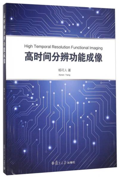
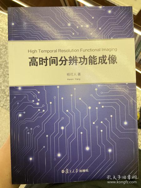
高时间分辨功能成像(英文版)
出版时间:
2016-05
版次:
1
ISBN:
9787309122060
定价:
49.00
装帧:
平装
开本:
16开
纸张:
胶版纸
页数:
186页
字数:
278千字
正文语种:
英语
-
20世纪70年代发现核磁共振(MRI)以来,其应用在近30年间得到飞速发展,目前它已成为现代社会无伤害性医学成像技术中重要的技术之一。随着神经科学的迅速发展,20世纪90年代初功能性核磁共振(fMRI)被引入,并成为当今探索大脑活动有价值的工具之一。《高时间分辨功能成像(英文版)》详细介绍了核磁共振、功能性核磁共振的成像原理及应用方法。
成像的速度是核磁共振重要的衡量指标,Multiband(MB)成像是前沿的高时间分辨率成像技术,可用于同时采集多层图像。《高时间分辨功能成像(英文版)》将介绍Multiband成像原理及其3个应用,具体包括静态功能性核磁共振(resting-state fMRI)、躯体感觉区功能性核磁共振(somatosensory fMRI)和磁共振动脉自旋标(ASL)。 杨可人,英国诺丁汉大学医学物理系博士。博士期间曾获得包括2003年诺贝尔生理与医学奖获得者曼斯菲尔德爵士等多年指导。在静态功能性核磁共振、高时间分辨率成像方向有多年研究经验。多次参加行业学术研讨会(ISMRM)并发表演讲。 Abstract
Acknowledgements
1 Introduction
1.1 Comparison between the MRI and others
1.2 Overview of the book
2 Nuclear magnetic resonance and magnetic resonance imaging
2.1 NMR
2.1.1 Introduction
2.1.2 Nuclei and spin, energy levels
2.1.3 Spins in magnetic eld
2.1.4 Boltzmann statistics
2.1.5 Rotating frame
2.1.6 Relaxation
2.1.7 Bloch equation
2.1.8 Chemical shift
2.2 MRI
2.2.1 Introduction
2.2.2 Gradients
2.2.3 Slice selection
2.2.4 Spatial encoding
2.2.5 k-space
2.2.6 Gradient echo imaging
2.2.7 Spin echo imaging
2.2.8 Echo planar imaging
2.2.9 Turbo spin echo imaging
2.2.10 GRASE
2.2.11 Parallel imaging
2.2.12 Simulation of matrix inversion
2.3 Hardware
2.3.1 Overview
2.3.2 Main magnet
2.3.3 Shim coils
2.3.4 Gradient coils
2.3.5 RF system
Bibliography
3 Functional magnetic resonance imaging
3.1 Introduction
3.2 Cerebral cortex
3.2.1 Somatosensory cortex
3.2.2 Motor cortex
3.2.3 Visual cortex
3.3 BOLD contrast
3.4 fMRI data acquisition
3.5 Paradigm
3.6 Data analysis
3.6.1 Preprocessing
3.6.2 Statistical analysis
3.7 Cortical flattening
3.8 Resting state
3.9 Perfusion imaging
3.10 Conclusion
Bibliography
4 A comparison of resting state functional connectivity
using fast imaging: 2D GEEPI, 3D GEEPI and 3D PRESTO
4.1 Introduction
4.1.1 Brain networks
4.1.2 Default mode network (DMN)
4.1.3 Sensorimotor network
4.1.4 Visual network
4.1.5 Dorsal attention network
4.2 Overview of fast imaging sequences
4.2.1 Echo planar imaging
4.2.2 PRESTO
4.3 Methods
4.3.1 Data acquisition
4.3.2 Data analysis
4.4 Results
4.4.1 No physiological noise correction
4.4.2 Post physiological correction
4.5 Discussion
4.6 Conclusion
Bibliography
5 Implementation and application of multiband echo planar
imaging in conjunction with SENSE
5.1 Introduction
5.2 Multiband imaging
5.2.1 Introduction
5.2.2 Multiband RF pulses
5.2.3 Optimization
5.2.4 Reconstruction
5.3 Implementation
5.4 Applications
5.4.1 Resting state fMRI
5.4.2 Somatotopic mapping of both primary and secondary somatosensory cortex
5.4.3 Multiband DABS ASL
5.5 Conclusion
Bibliography
6 Orientation mapping at ultra-high field
6.1 Visual cortex
6.2 Retinotopic organization
6.3 Orientation column organization
6.4 Introduction to the study
6.5 Methods
6.5.1 Stimuli and paradigm
6.5.2 fMRI data acquisition
6.5.3 Pre-processing
6.5.4 Data analysis
6.6 Results
6.7 Discussion
6.8 Conclusion
Bibliography
Abbreviations
Index
-
内容简介:
20世纪70年代发现核磁共振(MRI)以来,其应用在近30年间得到飞速发展,目前它已成为现代社会无伤害性医学成像技术中重要的技术之一。随着神经科学的迅速发展,20世纪90年代初功能性核磁共振(fMRI)被引入,并成为当今探索大脑活动有价值的工具之一。《高时间分辨功能成像(英文版)》详细介绍了核磁共振、功能性核磁共振的成像原理及应用方法。
成像的速度是核磁共振重要的衡量指标,Multiband(MB)成像是前沿的高时间分辨率成像技术,可用于同时采集多层图像。《高时间分辨功能成像(英文版)》将介绍Multiband成像原理及其3个应用,具体包括静态功能性核磁共振(resting-state fMRI)、躯体感觉区功能性核磁共振(somatosensory fMRI)和磁共振动脉自旋标(ASL)。
-
作者简介:
杨可人,英国诺丁汉大学医学物理系博士。博士期间曾获得包括2003年诺贝尔生理与医学奖获得者曼斯菲尔德爵士等多年指导。在静态功能性核磁共振、高时间分辨率成像方向有多年研究经验。多次参加行业学术研讨会(ISMRM)并发表演讲。
-
目录:
Abstract
Acknowledgements
1 Introduction
1.1 Comparison between the MRI and others
1.2 Overview of the book
2 Nuclear magnetic resonance and magnetic resonance imaging
2.1 NMR
2.1.1 Introduction
2.1.2 Nuclei and spin, energy levels
2.1.3 Spins in magnetic eld
2.1.4 Boltzmann statistics
2.1.5 Rotating frame
2.1.6 Relaxation
2.1.7 Bloch equation
2.1.8 Chemical shift
2.2 MRI
2.2.1 Introduction
2.2.2 Gradients
2.2.3 Slice selection
2.2.4 Spatial encoding
2.2.5 k-space
2.2.6 Gradient echo imaging
2.2.7 Spin echo imaging
2.2.8 Echo planar imaging
2.2.9 Turbo spin echo imaging
2.2.10 GRASE
2.2.11 Parallel imaging
2.2.12 Simulation of matrix inversion
2.3 Hardware
2.3.1 Overview
2.3.2 Main magnet
2.3.3 Shim coils
2.3.4 Gradient coils
2.3.5 RF system
Bibliography
3 Functional magnetic resonance imaging
3.1 Introduction
3.2 Cerebral cortex
3.2.1 Somatosensory cortex
3.2.2 Motor cortex
3.2.3 Visual cortex
3.3 BOLD contrast
3.4 fMRI data acquisition
3.5 Paradigm
3.6 Data analysis
3.6.1 Preprocessing
3.6.2 Statistical analysis
3.7 Cortical flattening
3.8 Resting state
3.9 Perfusion imaging
3.10 Conclusion
Bibliography
4 A comparison of resting state functional connectivity
using fast imaging: 2D GEEPI, 3D GEEPI and 3D PRESTO
4.1 Introduction
4.1.1 Brain networks
4.1.2 Default mode network (DMN)
4.1.3 Sensorimotor network
4.1.4 Visual network
4.1.5 Dorsal attention network
4.2 Overview of fast imaging sequences
4.2.1 Echo planar imaging
4.2.2 PRESTO
4.3 Methods
4.3.1 Data acquisition
4.3.2 Data analysis
4.4 Results
4.4.1 No physiological noise correction
4.4.2 Post physiological correction
4.5 Discussion
4.6 Conclusion
Bibliography
5 Implementation and application of multiband echo planar
imaging in conjunction with SENSE
5.1 Introduction
5.2 Multiband imaging
5.2.1 Introduction
5.2.2 Multiband RF pulses
5.2.3 Optimization
5.2.4 Reconstruction
5.3 Implementation
5.4 Applications
5.4.1 Resting state fMRI
5.4.2 Somatotopic mapping of both primary and secondary somatosensory cortex
5.4.3 Multiband DABS ASL
5.5 Conclusion
Bibliography
6 Orientation mapping at ultra-high field
6.1 Visual cortex
6.2 Retinotopic organization
6.3 Orientation column organization
6.4 Introduction to the study
6.5 Methods
6.5.1 Stimuli and paradigm
6.5.2 fMRI data acquisition
6.5.3 Pre-processing
6.5.4 Data analysis
6.6 Results
6.7 Discussion
6.8 Conclusion
Bibliography
Abbreviations
Index
查看详情
-
八五品
河北省保定市
平均发货23小时
成功完成率94.18%
-
九五品
海南省海口市
平均发货14小时
成功完成率84.33%

 占位居中
占位居中

